
Long Beach Animal Hospital offers canine laser neuter services for dog owners in Southern CA
One of the most common surgical procedures we perform is a dog neuter, know medically as an orchectomy. It is performed for several reasons:
- It minimizes roaming
- It minimizes aggressive behavior
- It prevents male dogs from impregnating females
- It prevents most prostate problems
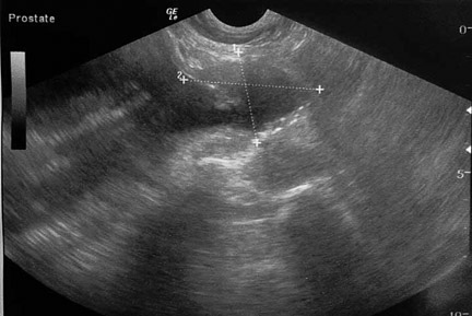
This is what the prostate gland looks during ultrasound
At the Long Beach Animal Hospital use of the laster is mandatory for all neuters. In this page we will first show you the surgery using the laser, then the traditional way this surgery is performed without the laser. The advantages of using the laser will be obvious.
Sometimes people get a jaded mindset when it comes to routine surgeries like neuters, that are performed by the thousands, especially at low cost spay and neuter clinics. It is a major surgery, and we treat it as such at the Long Beach Animal Hospital, which you will learn about in this page.
Several days prior to any surgery please bring in your pet for a preanesthetic exam and blood panel to confirm your pet is ready for anesthesia. At that time one of our doctors will go over any questions you have.
On the day of surgery we need your dog in the hospital between 7:30 AM and 8 AM. Please take away all food when you go to bed the evening before surgery. Let your pet have water during the night. Do not give your dog anything to eat or drink the morning of surgery.
Our surgeon will call you after the surgery is complete and your dog is awake. It can go home in the late afternoon the day of surgery unless instructed otherwise. Please call our office at 4 PM for pickup time, you will be given written post operative instructions then. We are open until midnight if you need to pick up later.
This page shows the surgical procedures for:
- A normal testicle using the laser
- A short video of a bloodless laser neuter at our hospital
- A testicle that has not completely descended into the scrotum and is in the inguinal canal using the scalpel blade
- A testicle that is still in the abdomen using the scalpe blade
- The use of neuticles
The following contains graphic pictures of an actual surgical procedure performed at the hospital.
Anesthesia
Pre-anesthetic preparation is important in every surgery we perform, no matter how routine. Surgery is not an area to cut corners. All of our neuters receive a physical exam prior to surgery.
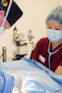
The pre-surgery physical exam is performed before any anesthesia by the surgeon doing the neuter that day
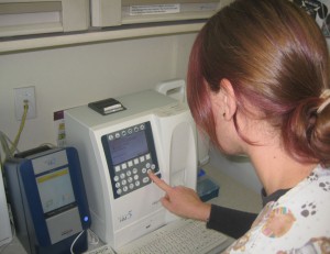
We prefer to take the pre-anesthetic blood panel one week prior to surgery. If we haven’t already taken a blood panel prior to surgery, we can take one the day of surgery and have a report within 30 minutes
Once a pet is anesthetized, prepared for surgery, and had its monitoring equipment hooked up and reading accurately, the surgery can begin.
We keep a close tab on important physiologic parameters for all of our surgeries. Monitors like this give us an early warning of an impending problem. Our patients are carefully monitored to detect any abnormalities before they become a problem.
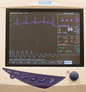
This machine monitors:
Temperature
Heart Rate
Heart rhythm
Oxygen saturation
Carbon dioxide level
Respiratory rate
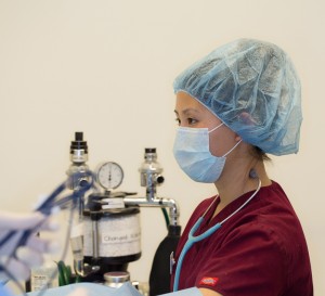
In addition to our monitoring equipment, our anesthetist stays “hands on” in monitoring important physiologic parameters.
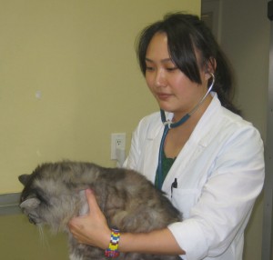
Our anesthetist is checking this pets mucous membranes to make sure enough oxygen is in the cardiovascular system
Even though our monitor check the heart rate and shows the EKG, our anesthetist checks the heart with a stethoscope periodically throughout the procedure
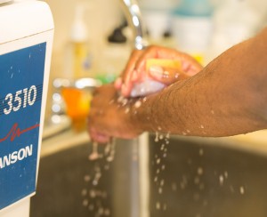
Once our surgeon has scrubbed up and is in a sterile gown, gloves, and mask, the surgery begins
While our patient is being anesthetized our surgeon is already in our surgical suite setting up instruments. Our surgeon is ready to start before our patient is at a proper plane of anesthesia. Once the anesthetist gives the green light the surgery starts immediately. We want our surgeon ready and waiting for his patient, not the other way around. All of this is to minimize anesthetic time.
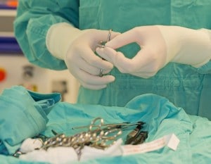
Sterile instruments are used on every surgery

Our surgeon does one final check on your pet’s stability and level of anesthesia before beginning the procedure
Laser Neuter Surgery
Using the laser has many advantages over using a scalpel blade. These include negligible bleeding during the procedure and minimal to no post operative pain.
The laser is calibrated specifically for each individual surgery
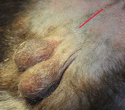
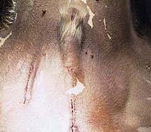
This dog is laying on its back, with its head towards the right. The red line show where the prescrotal neuter incision will be
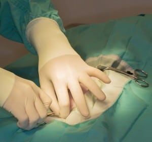
Once our patient is draped the procedure can begin
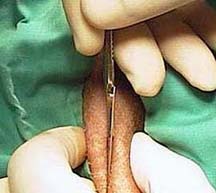
Surgery starts with our surgeon gently palpating the testicles and make the laser incision just in front of them in the prescrotal area (red line above)

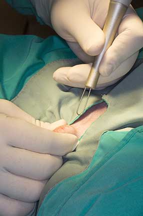
The initial skin incision in the skin just in front of the scrotum

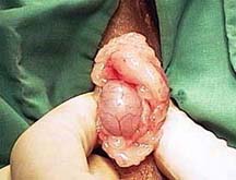
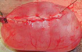
Notice the lack of bleeding from the skin as we enlarge the incision for room to remove the testicle. The white structure in the center is the testicle covered with protective tissue.


From this video you can see the lack of bleeding
Video of another dog being neutered with the laser

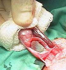
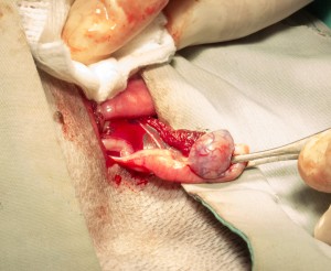
All of the pertinent structures associated with the testicle are apparent in this picture. The white horizontal line below the surgeon’s thumb is the epididymus. The dark structure to the right of this is the panpiniform plexus. This plexus contains arteries and veins to cool the testicle, supply it with nutrients, and allow testosterone to go into the bloodstream.


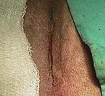
The appearance of the blood-free incision just prior to suturing the skin
The final appearance of the sutured skin, with no swelling or bleeding.
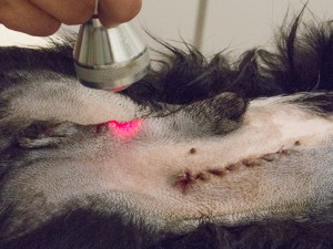
In addition to using the carbon dioxide laser to perform the surgery, we use the companion laser to aid the healing process at the incision before our patient is fully awake. This decreases post operative swelling so your pet is much more comfortable. When combined with the actual medication for pain we administer, our patient wakes up with minimal to no pain, and the healing process progresses faster.

Companion Laser in use immediately after a laser neuter
Click on the video to see it in action after a neuter surgery
Most dogs recover from this laser neuter surgery by the next day. It is important to keep these dogs quiet for a few days postoperatively to allow the incision sites to heal. In most neuters we put in sutures that are just under the skin and dissolve on their own, so there is no need to return for suture removal. If sutures are placed in the skin, we usually remove them in 7-14 days.
Our Laser Page has detailed information on the use of the laser for various other surgeries besides neuters.
Cryptorchid surgery
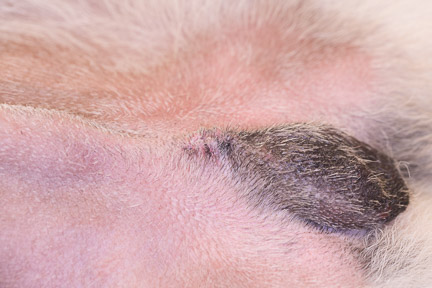
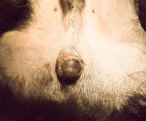
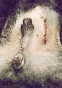
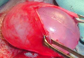
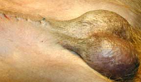
The dog being neutered in this picture has only one testicle in the scrotum, called a cryptorchid, monorchid, or retained testicle. This happens in only a small percentage of the animals we neuter. The other testicle can be in the abdomen or in the inguinal canal (inner thigh region).

In this dog’s case it is in the inguinal canal, as evidenced by the bulge (arrow). It is important to remove the retained testicle because it can become cancerous later in life.
First we will remove the normal testicle in the scrotum with a prescrotal incision you have already seen above. Since our patient in this case has one of his testicles in the inguinal area, the surgery is not finished yet. The skin incision and exposure of the other testicle is similar to the normal testicle removal.
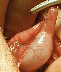
In the inguinal area there is significant fat under the skin.

The actual structures of the testicle and its blood supply are exposed, ligated with two sutures, and placed back into the inguinal area.

The incision is closed in two layers. First the subcutaneous tissue, then the skin.
Here is a final view of our patient and his two incisions. As he wakes up from anesthesia he will be given an injection for pain.
When the retained testicle is not in the inguinal area it is located in the abdomen. This testicle can also become cancerous so it is important to remove it.
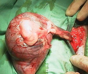
This patient only has one testicle in the scrotum. The other is in the abdomen.
In this picture the testicle has been brought out through the inch incision in the abdomen
A closeup of this testicle, which is smaller (atrophied) compared to the one we removed from the scrotum
The post operative appearance showing both incision sites, with the abdominal incision on the right
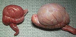
You can easily see the difference in size between the atrophied testicle in the abdomen (left) and the normal testicle from the scrotum on the right. The testicle in the abdomen was removed at a young age so it never had a chance to become cancerous.
This is the appearance of a different dog that had a cancerous testicle, called a seminoma. It was not removed until later in life, so it had a chance to enlarge tremendously.
Both incisions are treated with our companion laser just after surgery just like we do when both testicles are in the scrotum
Neuticles
An interesting variation on this neuter surgery is the placement of solid silicone implants in place of testicles. This gives a natural look after neuter surgery that is desirable to some people. We highly frown upon having this done, have only performed it once decades ago on a special case, and don’t plan on doing it again.

This is the appearance of a normal dog scrotum prior to neuter surgery. Use it as a basis of comparison at the end of this section to see what the scrotum looks like when we have implanted neuticles in place of the testicles.
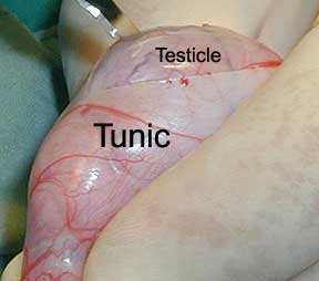
In this surgery the testicle is removed and the neuticle is placed in the sack that holds the testicle, called the tunic.
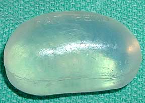
This is the sterile neuticle on the surgery tray ready for placement. Neuticles come in various sizes and shapes to be custom fitted to each individual.
The neuticle is gently implanted in place of the testicle. A proper fit is imperative, so it is important to order the proper size ahead of time.
When we are sure of a proper fit we carefully suture the tunic with a suture material that will eventually dissolve
This is the final appearance after the placement of the neuticles