
Introduction
Ferret’s are highly prone to several serious diseases. In addition to insulinomas and adrenal gland disease, they are also prone to cancer. This page shows a ferret that has a liver cancer.
What is Cancer?
The scientific word for cancer is neoplasia, meaning new growth. In reality, it is an abnormal growth of cells that interferes with an organ’s ability to function, resulting in a degree of failure in that organ.
Some of these abnormal cells break off from the organ and spread to other organs in the body, causing them to fail. This process is called metastasis, and is the hallmark of malignant cancer.
To learn much more about cancer in animals follow this link.
What Causes Cancer in Ferrets
Since cancer is prevalent in ferrets, in addition to the usual environmental and hormonal factors that are associated with cancer, there seems to be some type of genetic predisposition.
Symptoms of Cancer in Ferrets
Some ferrets do not show any symptoms while others exhibit lethargy and weakness and a distended abdomen. There might be a decrease in appetite and weight loss. Symptoms alone are not enough to diagnose liver cancer.
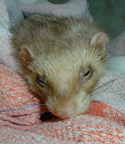
It is easy to see this is an ill ferret
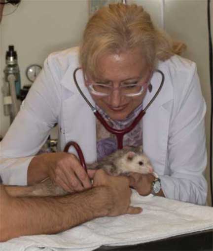
The first order of business for any ill ferret presented to the LBAH is a physical exam
Upon admission to our hospital any ill ferret has its blood glucose checked and it’s temperature taken due to their propensity to hypoglycemia and hypothermia (low blood sugar and low body temperature)

The first thing we do with a sick ferret is take its body temperature
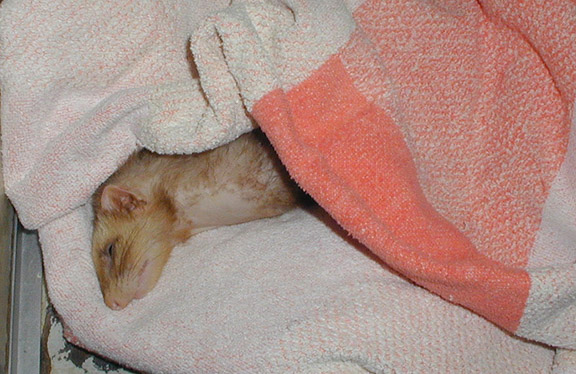
If low we immediately warm them up with warm towels

Next we check the blood glucose level using an instrument that only requires a few drops of blood
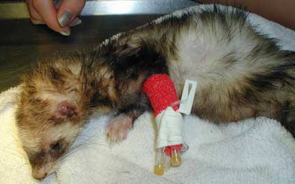
Once we assess those two parameters we put in an IV (intravenous catheter) and administer fluids
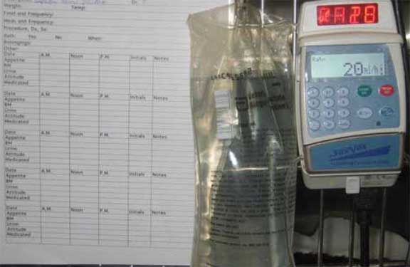
A special type of fluids are given with an IV pump for accurate dosing
Fluids are a crucial part of most every treatment we do for a wide variety of animal that are ill. We have a complete page dedicate to this important treatment. Click here to learn much more.
We also monitor the blood pressure and heart rate to determine if any other therapy is needed
Diagnosis of Cancer in Ferrets
Like most diseases, the diagnosis of liver cancer is made based on history, examination findings, and diagnostic tests. This is what is called the Diagnostic Process, and you can learn more about it here.
Blood Panel
Blood samples are an aid in the diagnosis and can also be helpful to rule out other important diseases that might be occurring simultaneously. Our laboratory specializes in working with ferrets, and has a special blood panel for them.
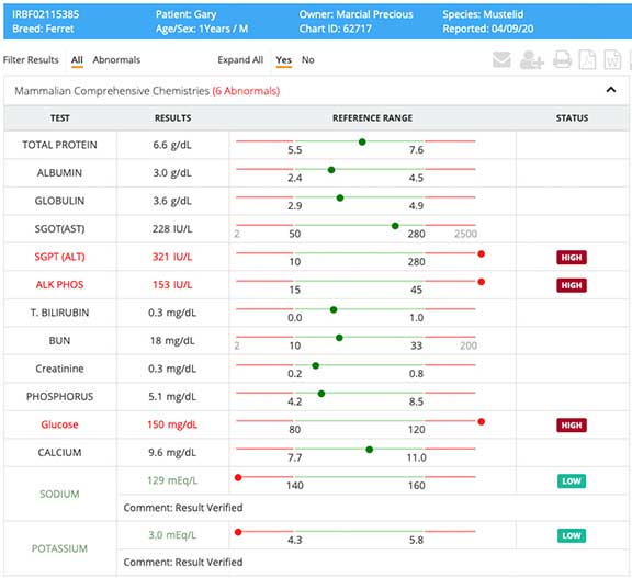
This ferret blood panel shows elevated liver enzymes (ALT and Alk Phos), a common finding in liver cancer
Radiology
A useful tool in diagnosing cancer in a ferret is radiography. It gives us a clue as to the presence of fluid along with abnormalities of the internal organs
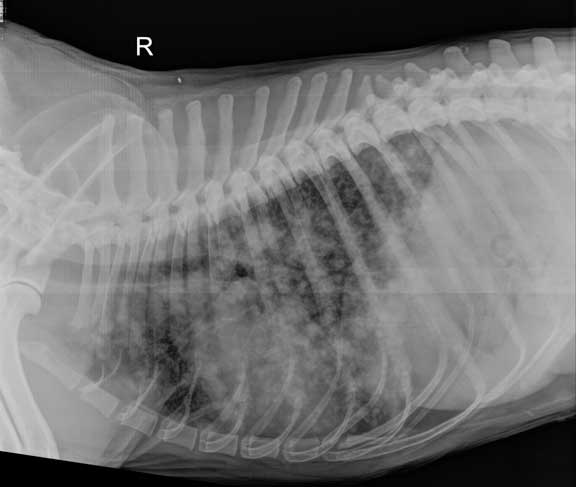
This ferret is laying on its right side, and has a distended abdomen due to a large liver and fluid in its abdomen. It also has a large spleen.

In addition, there are 3 metallic clips visible just under the kidney from a prior surgery for adrenal gland disease. The clips were used to stop bleeding when the diseased adrenal gland was removed. The identified organs are: Liver (L), Stomach (S), Kidney (K), Spleen (SP) and Bladder (B).

A view of this same ferret while it is on its back shows the large area of the liver and the large spleen. Look carefully at the spleen because it is folded over on itself in this picture and is larger than it first appears.

This test tube is filled with fluid that was taken from a ferret that had a distended abdomen due to liver cancer. Analysis of this fluid in conjunction with other diagnostic tests gives us a clue as to its cause.
Whenever we suspect cancer we take a radiograph of the chest to look for evidence that the cancer has spread, called metastasis. We use the chest for this radiograph because the small blood vessels that are located around the lungs act as a filter to trip cancer cells that have spread. The black background of the lungs allows us to visualize these cells that are white in color.
All this white splotches are cancer that has spread to the lungs in this pet
Ultrasound
Ultrasound is a valuable tool in refining the diagnostic once a problem is identified on a radiography. It is such a valuable tool that we have dedicate a full page to it. Click here to learn more.
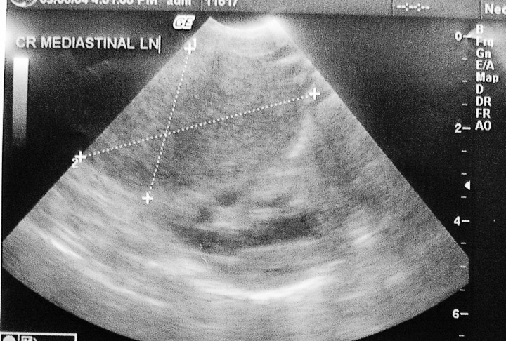
This is what an enlarged lymph node in the chest looks like during ultrasound
Treatment Decision
In many cases of cancer in ferrets the cancer has invaded an internal organ and surgery is needed. Since ferrets are prone to so many internal organ diseases, and since exploratory surgeries can be performed rapidly and yield a large amount of information, it is common to perform surgery instead of ultrasound.
Some ferrets are diagnosed as having cancer when an abdominal surgery is being performed for other reasons (adrenal gland disease, spay, insulinoma). This is especially true for the ferrets that are not showing any symptoms of this disease.
Pre-surgical Preparation
Once the decision has been made to perform surgery we start the preparation long before the surgical procedure.
This is a sterile abdominal surgery, and our surgeon starts the pre-surgical process by using special soap to clean his hands.

We use a special surgical scrub and brush to scrub before putting on sterile gloves
While our patient is being anesthetized our surgeon is already in our surgical suite setting up instruments. Our surgeon is ready to start before our patient is at a proper plane of anesthesia. Once the anesthetist gives the green light the surgery starts immediately. We want our surgeon waiting for his patient, not the other way around. All of this is to minimize anesthetic time.

Our doctor waiting patiently prior to surgery

While waiting instruments are prepared so that the surgery can start as soon as the patient is brought into the surgical suite
Anesthesia
If it is determined that surgery is needed for a ferret with cancer will have a specific anesthetic protocol. When everything is to our satisfaction we will administer a sedative. This will calm the pet down and make the administration of the actual anesthetic, along with post operative recovery, much smoother.
Once a pet is anesthetized, prepared for surgery, and had its monitoring equipment hooked up and reading accurately, the surgery can begin.
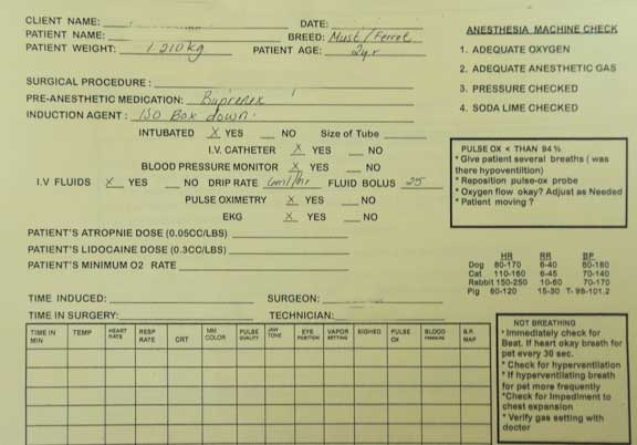
We use a detailed anesthetic form for every surgery
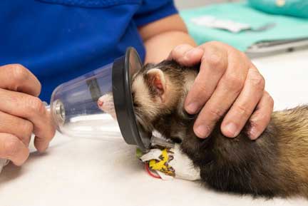
We initially administer anesthetic to our sedated pet via face mask
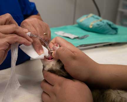
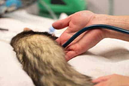
All our ferret surgery patients are then intubated for accurate administration of anesthesia and oxygen. You saw where this tube goes in the chest radiograph earlier in this page.

Our surgeon and anesthetist work closely together
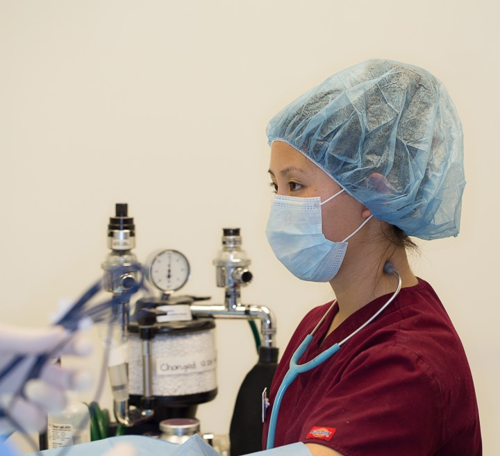
We use a special stethoscope (called an esophageal stethoscope) that is passed down the esophagus and can give us a clear sound of the heart
We keep a close tab on important physiologic parameters for all of our surgeries. Monitors like this give us an early warning of an impending problem.

This machine monitors:
Temperature
Heart Rate
Heart rhythm
Oxygen saturation
Carbon dioxide level
Respiratory rate
We also carefully monitor oxygen saturation on our surgical patient with a special and small pulse oximeter
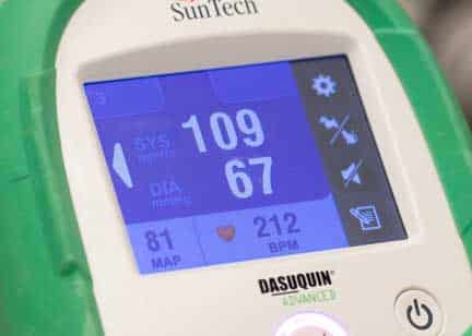
We constantly monitor blood pressure and heart rate. This ferret has a blood pressure of 109/67, and a heart rate of 212 beats per minute. This is normal for a ferret under anesthesia.
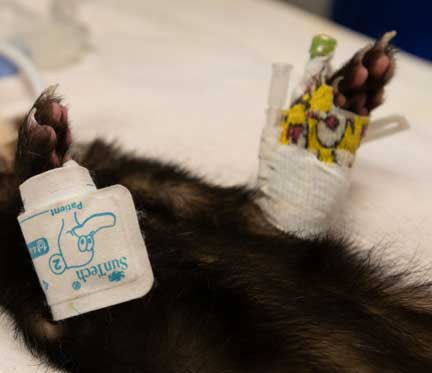
You can see the pediatric blood pressure probe on the right leg, and the IV catheter on the left leg, of this ferret under anesthesia
In addition to our monitoring equipment our anesthetist stays “hands on” in monitoring important physiologic parameters.
Our anesthetist has access to the patient at all times before, during, and after the procedure
Surgical Treatment
When the signalment, history, physical exam, and diagnostic tests all indicate a cancer of one of the internal organs, surgery is indicated in many cases.
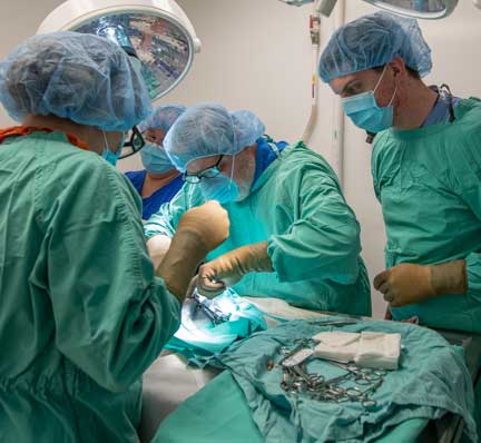
In cancer surgery it is very common for our surgical team to consist of 2 doctors and one student
Several precautions must be taken when this surgery is performed. Ferrets can easily become hypothermic due to the anesthesia and the fact that their abdomen will be open during the procedure. They can also become hypoglycemic due to the stress of the procedure. Special precautions are taken to help mitigate these problems.
This ferret is a high risk anesthetic patient. In addition to the usual precautions we take for hypoglycemia and hypothermia, we have to be concerned with the induction of anesthesia due to a weakened condition.

Our patient is carefully clipped and scrubbed
Our surgeon does the all important draping after the final cleanse of the abdomen

Under these drapes is a hot water blanket and hot water bottles to prevent hypothermia

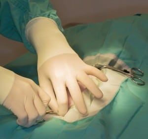
The initial incision is made on the midline of the abdomen

This midline incision allows access to an important landmark called the linea alba. This is a tendinous attachment between the abdominal muscles, and is the area where we put our sutures into the abdomen at the end of the surgery to prevent a hernia of abdominal organs.

After the linea alb is opened with a scalpel, a scissor is used to extend the incision and give full access to the abdomen
On a pet with cancer we sometimes encounter an abdomen that is filled with fluid or blood. This is usually suctioned away to visualize the internal organs.
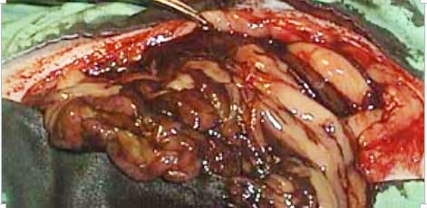
This unsightly picture gives you an idea of what we might encounter once the abdomen is open
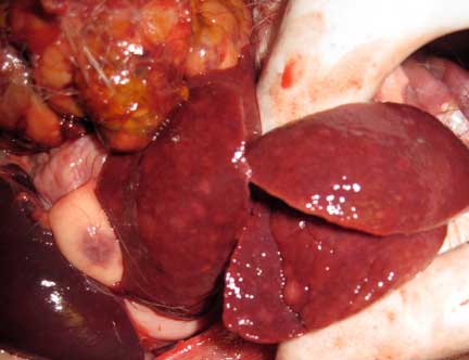
This is a normal looking liver with the gall bladder (it stores bile) underneath in the middle
A full view of the gall bladder with the liver lobes retracted
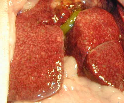
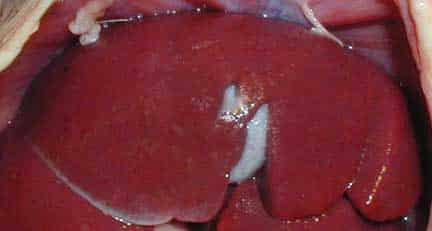
This is an unhealthy liver that could have hepatitis or cancer. Compare it to the liver picture above of a healthy liver.
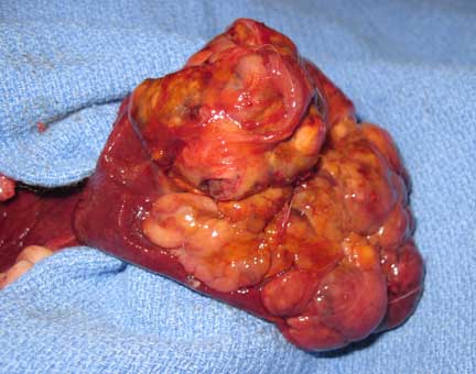
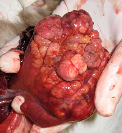
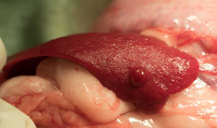
This liver lobe that is filled with cancer has been exteriorized from the abdomen and is laying on a surgical drape. Not the healthy liver lobe on the left part of this cancer filled liver lobe.
This lobe of cancerous liver in the surgeons hands just before removing it
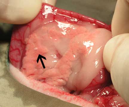
The other liver lobes (you can see the cancerous lobe at the top left) are not healthy. They have nodules in them that are probably from the spread of the cancer from the cancerous liver lobe at the top left. A liver in this condition is a poor prognosis.
Once we are done with the liver part of our surgery we check all of the other abdominal organs. This is a ferret spleen with a nodule on it that is probably not causing any pathology.
This is a pancreas with an insulinoma nodule. You can learn more about ferret insulinoma’s at this link
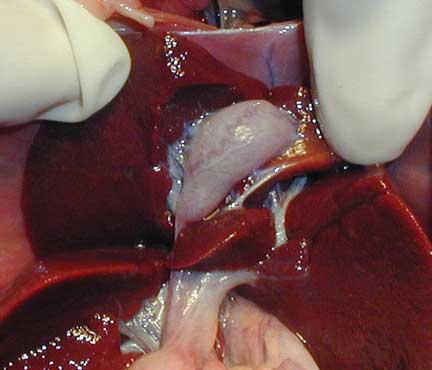
A very important organ to check during and exploratory surgery is the adrenal gland since adrenal gland disease is common in ferrets.
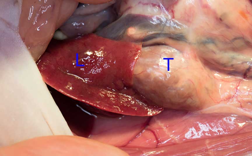
This is an adrenal gland tumor (T), under a lobe of a healthy liver (L)
Prognosis for a Ferret with Liver Cancer
Animals in general have strong survival instincts that helps them in hiding disease externally when on the inside they are quite ill. This makes it hard for owners to see any initial symptoms of cancer like a change in appetite or weight loss until it has progressed to the point that the disease has spread. This leads to a poor prognosis in many cases.
This emphasizes the importance of a yearly (and more often as you pet ages) wellness exams. Our pets do not live as long as we do, and disease problems occur sooner than we expect. This exam we perform while your ferret is apparently healthy is called a Wellness Exam. This applies to all pets in addition to animals like ferrets the are highly prone to many serious diseases.
Our doctors will also show you how to do an In-Home-Exam so you can identify potential problems early and bring your pet in for a proper diagnosis early in the course of a disease when the prognosis for treatment is much better.
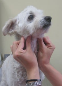
As a part of teaching you this exam you get to check your pets lymph nodes. In this case the owner of this dog is learning how to palpate the submandibular lymph nodes.