
Introduction
Chinese Water Dragons are fun pets that require very specific environmental conditions. When these conditions are not met problems can occur, in this case the pet was unable to lay its eggs and was feeling ill. This page on Chinese Water Dragon Spay will give you all of the details.
You can learn more about our standard of care of sick reptiles from the Association of Reptile and Amphibian Veterinarians.
Because these eggs were becoming toxic it was decided to remove them in version of a surgery called an ovariohysterectomy (OVH), more commonly known as a spay. This surgery prevents this Water Dragon from laying eggs in the future, so it will not encounter this illness again.
They are cute little guys. This one has a broken forearm.
This page shows graphic pictures from an actual surgery.
Husbandry
These animals originate from Southeast Asia so they require high humidity with plenty of water and a temperature range of 82-97 degrees F. They are tree climbers so make sure their cage has plenty of branches.
They need large cages to feel secure. If the cage is too small they will constantly rub their noses to the point that they will rub the bone raw. Male Water Dragons are territorial, so only one male should be in a cage. Two females can be kept with each male.
Insects and other arthropods, along with small mice, fruits, and vegetables are good foods to give them. Always use a multipurpose vitamin powder in their food several times per week.
Reproduction
Approximately twice per year they lay 8-12 eggs. They should be incubated in vermiculite at 78-80 degrees F for approximately 3 months. Make sure no insects bother them while they are incubating.

This is what we normally want to see, eggs laid on vermiculite
In the case that follows the Water Dragon was unable to lay its eggs, which caused it to become ill. Surgical intervention was needed to correct the problem.
Diagnosis
A patient like this is not eating well and, a distended abdomen (called the coelomic cavity in birds and reptiles) can be palpated during an examination.

An x-ray reveals the extent of the eggs in our patient. The coelomic cavity (reptile equivalent of the abdomen) is filled with eggs.
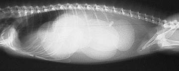
If you cannot see them in the above radiograph this might help
Click here if you want to learn more about Reptile X-rays.
Anesthesia
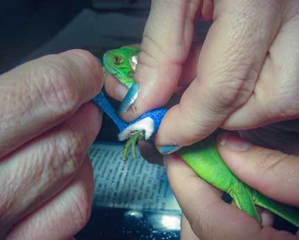
Anesthesia is very important, especially in such a small patient. To minimize the risk of anesthesia we insert a flexible breathing tube into the trachea (windpipe) of our patient. This tube allows us to give oxygen and anesthesia in very refined quantities. It also allows us to inflate the lungs since reptiles commonly do not breathe on their own when anesthetized.

It takes lots of fingers and good lighting to be able to gently put the small breathing tube into the trachea
After the breathing tube has been placed and our patient anesthetized it is prepared for surgery. We use several instruments to monitor the oxygen level of our patient during surgery, the primary one being a Pulse Oximeter. The Pulse Ox is an instrument that measures oxygen saturation in the red blood cells. It is instruments like this, along with our extensive expertise with reptiles, that allows us to safely anesthetize such a small patient.

This Pulse Ox is showing an oxygen saturation of 94% (normal) and a heart rate of 157 beats per minute. These are good numbers for a pet that is under anesthesia.
On the right side of this picture you can see a probe placed on its tail. This probe goes to the Pulse Ox.
We have much more information on how we anesthetize a wide variety of animals, ranging from a tiny bird to a 200 pound Great Dane. Click here to learn more about anesthesia.
Surgery
This is a sterile procedure, and we perform it in an aseptic manner just like any other surgery.
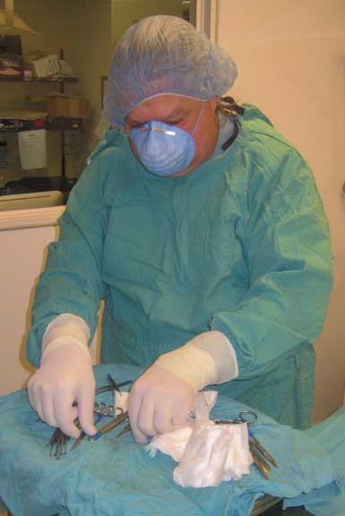
Our doctor for this surgery is fully capped, masked, and gloved. Dr. R our surgeon is sorting his sterile instruments just prior to starting the procedure.
Great care must be taken when making an incision into the coelomic cavity. There is a large vein that lies just under the scales, and if punctured, can cause extensive bleeding. Such a small animal can not tolerate blood loss that would be acceptable in other animals.
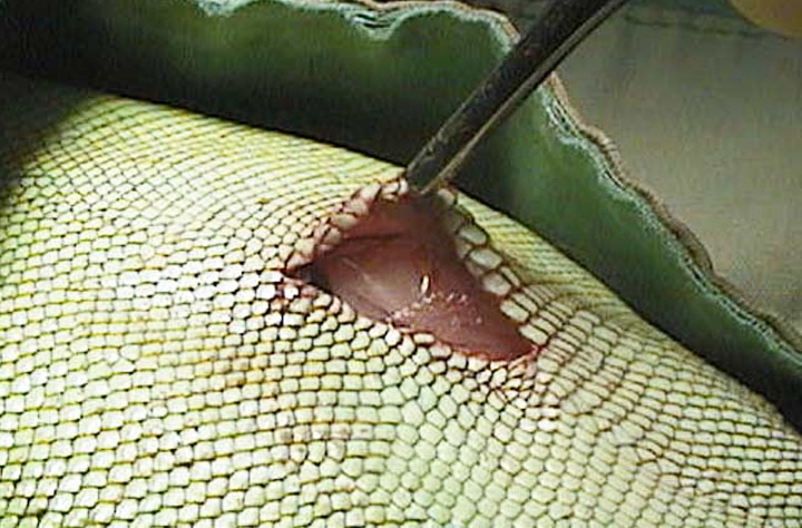
Lifting up the skin after the initial incision to identify the location of the vein
The coelomic cavity is filled with eggs that literally spill out when we make our incision. Each ovary with its associate eggs is isolated, and the shell gland with eggs is removed.

It is amazing how many eggs can be in this small creature
A special instrument called a hemoclip is used to clamp the blood vessels that lead to the ovary and shell gland. The hemoclip is used because it minimizes surgical time, so there is less risk of anesthesia. This instrument uses a small metal clip to stop the blood flow. The clip can be vividly seen on an x-ray because it is metallic.

Gently applying the hemoclip on the blood vessels that supply the ovary
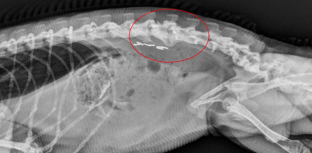
The metallic hemoclips are under the red circle of this iguana with arthritis called spondylosis
After surgery our patient is taken to a warm room at 85 degrees F for recovery. Pain medication is given, and temperature monitored, until it is well enough to go home.
As a fun comparison, we have pages on an Iguana Spay, Dog Spay, Rabbit Spay, and Cat Spay.