
These are close up views of the radiographs on the previous page. When you are done looking at them go back to the “What’s Your Diagnosis Page” to continue learning about radiography.
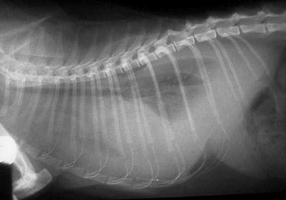
The thorax

W- windpipe (trachea)
H- heart
VC- vena cava
Sto- stomach
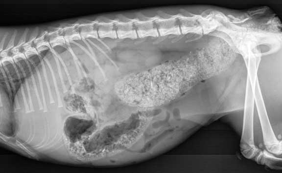
This view is towards the front of the abdomen

L- Liver
S- Stomach (filled with food)
K- Kidneys (they are overlapping)
LI- Large intestine (filled with stool)
SI- Small Intestine
Sp- Spleen
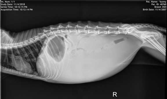
This view is towards the rear of the abdomen.

K- Kidneys (they are overlapping)
LI- Large intestine (filled with stool)
SI- Small Intestine
Sp- Spleen
B- Urinary Bladder (filled partly with urine)
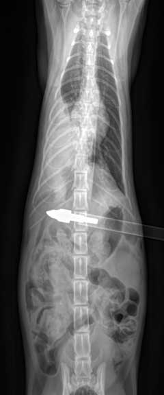
The white arrow points to stainless steel sutures from a spay (OVH).
Cat’s with Medical problems:
Side view of the abdomen showing severe fecal impaction in the colon called obstipation.
Fluid in the abdomen called ascites. Notice that you cannot see the individual internal organs.
Fluid in the chest called pleural effusion
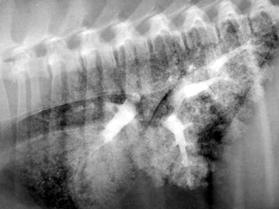
Barium in the lungs showing he extensive pulmonary vasculature

Pellet in the neck
Cat show with an arrow that penetrated the abdomen