
Introduction

Ferrets are prone to eating many things, especially when they are young. This s one of the way’s they explore their world, although it sometimes gets them into trouble (such is the nature of a ferret and makes them so endearing) and what they eat gets stuck in their gastrointestinal tract. This means it can be anywhere from the esophagus to the anus.
This page shows 2 different cases of ferrets with intestinal foreign bodies that need to be removed surgically, and how we diagnosed them. Let’s jump right into the diagnosis and show you a radiograph of the first one with the foreign body. This will be fun way to start this page and let you learn how to read a radiograph.
Before we show the radiograph you might want to review our How to Read a Radiograph page and brush up on some of those aspects of radiology you learned in veterinary school! Once you take a gander at that come on back here and interpret the following radiographs of the first ferret with the foreign body.

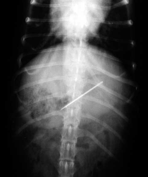
This ferret is lying on its back with the head at the top. It is called a VD (venture-dorsal) view
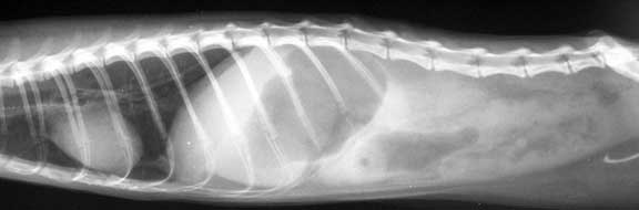
This ferret is lying on its right side, with the heat at the left
Good luck, the answer will be later in this page.
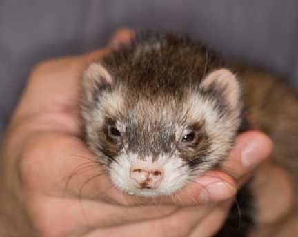
Ferret Behavior
These are inquisitive little critters, especially when young. They get into everything, so here is a list of things around your house you might want to make “ferret proof”:
Anything you drop on the kitchen floor
Smelly or tasty things you put in the garbage pale
Shiny objects like jewelry
Anything in the bathroom like toilet paper or tissue
Special care to minimize the chance of ingesting foods that are typically toxic to animals like chocolate and onions. Our Household Poisons page has a more complete list.
Be especially vigilant around the holidays with tinsel, gift wraps, and Christmas trees and ornaments. They might even drink the putrid water at the base of a Christmas tree, or bite into an electrical cord causing severe injury and even death due to pulmonary edema.
What is a foreign Body?
This is a general medical term for objects that don’t necessarily belong in the GI (gastro-intestinal) tract. Fortunately, many of them pass through the GI tract normally, and oftentimes the only indication that your pet ate something foreign is when they vomit it up (called emesis) or they pass it in their stool.
It might be hard to believe, but the needle in this dogs stomach passed through uneventfully
In the two ferret foreign body cases on this page the foreign bodies were not passing through, and needed to be removed surgically. Next, let’s learn how we make this diagnosis.
Diagnosis of an Intestinal Foreign Body in a Ferret
Making a diagnosis of a disease in an animal is not straight forward. They readily hide symptoms, do not cooperate during a physical exam, and have vastly different anatomies and physiologies than us humanoids. Then, add the fact that we are like pediatricians, and are trying to solve the medical puzzle through the eyes of a concerned (and sometimes not very concerned) father or mother.
Because of all of this, we have a methodical approach to making a diagnosis called the Diagnostic Process. This is another page you might want to read before coming back here to continue our saga of the ferret’s with intestinal foreign bodies.
The diagnosis process in animals involves the following 5 areas:
- Signalment
- History
- Physical Exam
- Diagnostic Tests
- Response to Treatment
Signalment
A young ferret that is presented with no appetite makes us think of an intestinal foreign body, an infection or trauma as the cause of this lack of appetite (called anorexia). This is opposed to the other common diseases of ferrets called Adrenal Gland Disease, Cancer, and Insulinoma, because they tend to occur in much older ferrets.
History
A history of not eating well (partial anorexia), or not eating at all (anorexia) is the first clue. Sometimes the ferret will be lethargic, and only on occasion will it be vomiting, as opposed to most other animals that vomit (emesis) when a foreign body is obstructing the GI tract.
Physical Exam
A ferret with a foreign body might have a perfectly normal physical exam and body temperature and weight. It might have a painful abdomen upon palpation, and depending on the size and location of the foreign body, we might be able to actually feel it.
If the foreign body has perforated the GI tract it can cause a peritonitis and sepsis. In this case the ferret is quite ill and needs immediate medical and surgical care.
Diagnostic Tests
On every sick pet we perform what is called a minimum database. This consists of the following:
Blood Panel- CBC
Routinely, unless the ferret is ill from a peritonitis or some other problem, the blood panel does not show any problems in most cases. If there is a problem it would tend to be an elevated WBC (white blood cell count) on the CBC.
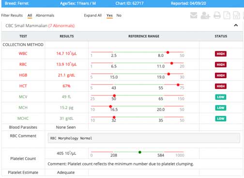
This is a ferret CBC
Blood Panel- Chemistry
The chemistry, sometimes called a BCP (Biochemistry Panel) analyses internal organs and does not look at the White and Red blood cells like a CBC.

This is what a ferret chemistry panel looks like
In the CBC and BCP panels above there are some abnormalities on the tests. In spite of these abnormalities, the ferret that this blood was taken from was normal. This shows the importance of interpreting diagnostic findings with the 4 other parameters of the Diagnostic Process.
Radiographs
This tends to be one of the more important diagnostic tests for an intestinal foreign body. Not every foreign body shows up readily on a radiograph. Those that are radiopaque are easy to spot, but those that radiolucent are not. Every foreign body is different, some are radiopaque, and some are radiolucent.
Here is the answer to the radiographs at the beginning of this page. The foreign body is circled.
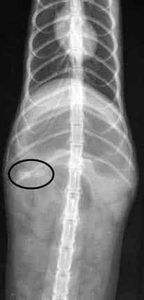
On the VD view the mildly opaque foreign body is at the beginning of the small intestines called the duodenum
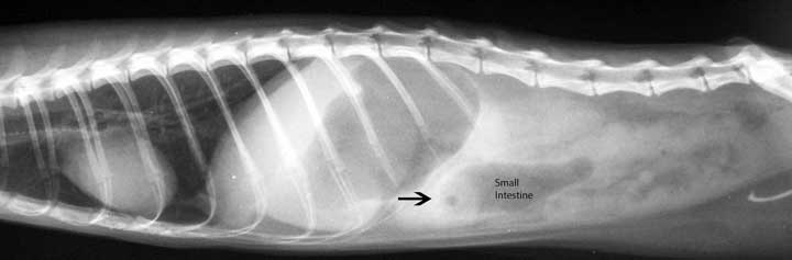
The arrow points to the circular FB on the lateral view in the same location. Note the dark area that says small intestine. This dark area is gas, because the foreign body is causing an ileus, and the intestine is filling up with gas and distend because the normal peristalsis is interrupted.
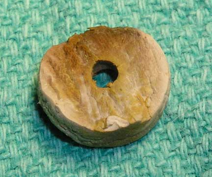
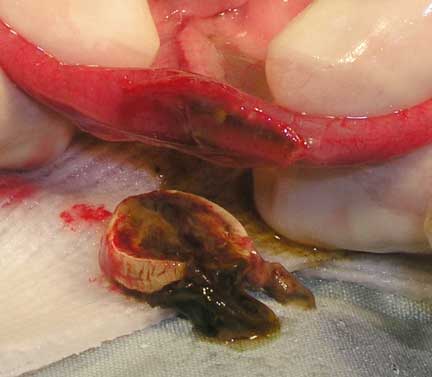
For a better understand here is that foreign body after it was removed surgically
Ultrasound
In the other intestinal foreign body case the FB was not seen radiographically and an ultrasound was performed to confirm the diagnosis.
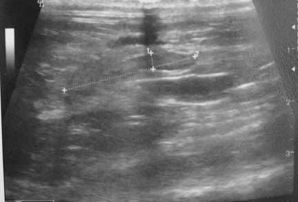
This is the ultrasound. The lines are measuring the size of it in the small intestine.
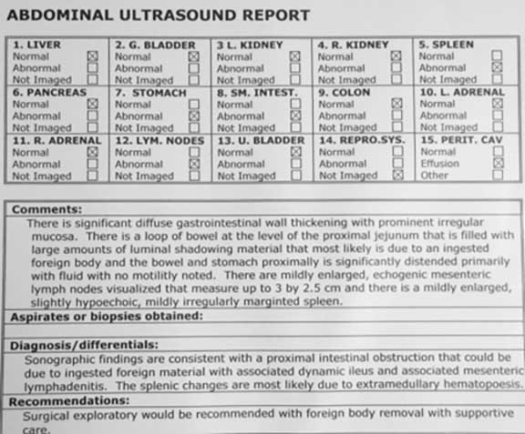
This is the report. Read the part from the “comments” on down
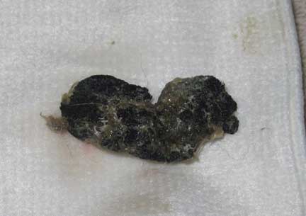
This is the plastic foreign body as seen in the ultrasound after it was surgically removed
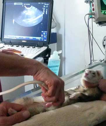
This is how we do an ultrasound on a ferret
Ultrasound has revolutionized veterinary medicine. In the “old days” before ultrasound we either had to give barium on a case like this, or just go on the Signalment, History, and Exam findings, and do an exploratory surgery and see if there was a FB. With ultrasound we now have a confirmed diagnosis, with the size and location of the FB in addition, to help plan surgery.
Doing ultrasound on an exotic type of animal like a ferret, as opposed to a dog or cat, is a special skill due to the tremendous amount of variability in anatomy. This is why we always call in a trusted radiologist named Ann Reed who was the necessary experience to do this.
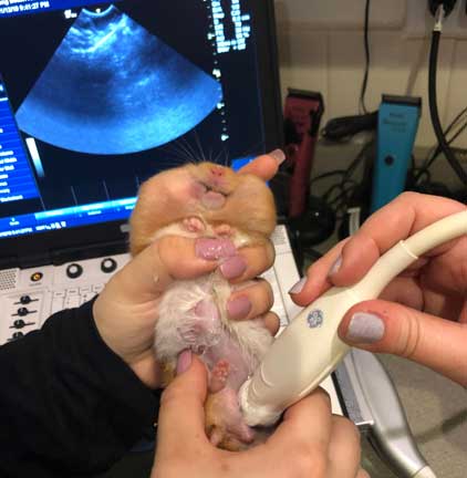
Here is Ann showing a veterinary student how to do an ultrasound on a hamster
Response to Treatment
If we made the correct diagnosis and implemented the proper medical and surgical therapy then the problem should be resolved. As to be expected, in both of these cases the ferrets returned to normal in a few days.
Let’s get to the surgery!
Surgery to Remove a Foreign Body in a Ferret
Pre-Surgical Preparation
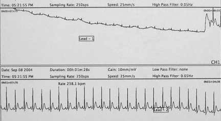
Once the blood panel, ultrasound and radiographs have been interrupted the next step is a pre-anesthetic EKG (also know as an ECG or electrocardiogram) to make sure there are no heart arrhythmia’s that would increase the risk of anesthesia.
Since ferrets are so prone to hypoglycemia (low blood sugar) and hypothermia (low body temperature ) extra precautions are taken on the day of surgery
The next step is one final exam and review of all records before any anesthetic is given
Surgeon Preparation
This is a sterile abdominal surgery, and our surgeon starts the pre-surgical process by using special soap to clean his hands.
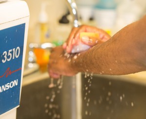
He washes his hands several times with the surgical soap and brush before putting on sterile gloves
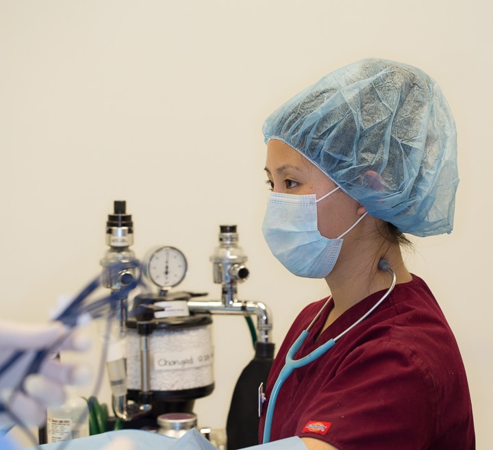
While our patient is being anesthetized our surgeon is already in our surgical suite setting up instruments. Our surgeon is ready to start before our patient is at a proper plane of anesthesia. Once the anesthetist gives the green light the surgery starts immediately. We want our surgeon waiting for his patient, not the other way around. All of this is to minimize anesthetic time.
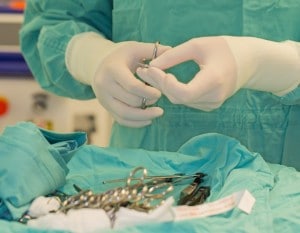
While our patient is being anesthetized our surgeon is already in our surgical suite setting up instruments.

Our surgeon is ready to start before our patient is completely prepped. Once the anesthetist gives the green light the surgery starts immediately. We want our surgeon waiting for his patient, not the other way around. All of this is to minimize anesthetic time.
Anesthesia
When everything is to our satisfaction we will administer a sedative. This will calm our patient down and make the administration of the actual anesthetic, along with post operative recovery, much smoother. Once a pet is anesthetized, prepared for surgery, and had its monitoring equipment hooked up and reading accurately, the surgery can begin.
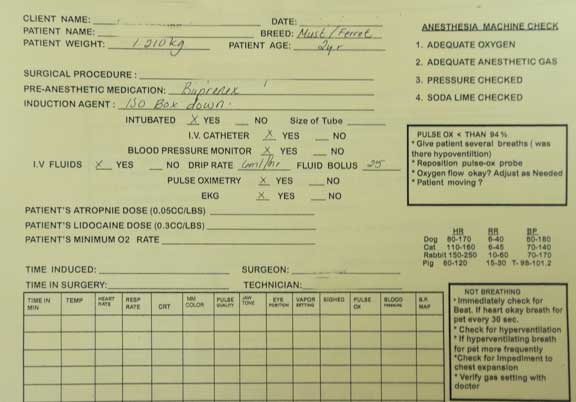
We use a detailed anesthetic form for every surgery
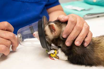
We initially administer anesthetic to our sedated pet via face mask
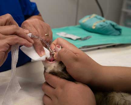
All our ferret surgery patients are then intubated for accurate administration of anesthesia and oxygen. You saw where this tube goes in the chest radiograph earlier in this page.
In ferret surgery pay particular attention to low body temperature (hypothermia) and low blood sugar (hypoglycemia). They are placed next to special hot water bottles throughout the procedure.

We keep a close tab on body temperature, before, during, and after surgery since they are prone to hypothermia
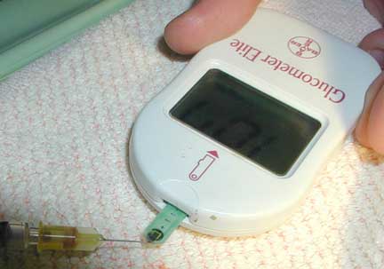
We also monitor the blood glucose levels during surgery since they are prone to hypoglycemia during the procedure
Our patients are carefully monitored to detect any abnormality before it becomes a problem. This early warning system is important in such a small animal that is ill and undergoing anesthesia and major surgery.

This machine monitors:
Temperature
Heart Rate
Heart rhythm
Oxygen saturation
Carbon dioxide level
Respiratory rate
We also carefully monitor the oxygen saturation of our surgical patients with a Pulse Oximeter
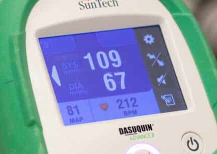
We constantly monitor blood pressure and heart rate. This ferret has a blood pressure of 109/67, and a heart rate of 212 beats per minute. This is normal for a ferret under anesthesia.
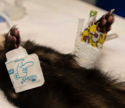
You can see the pediatric blood pressure probe on the right leg, and the IV catheter on the left leg, of this ferret under anesthesia
In addition to our monitoring equipment our anesthetist stays “hands on” in monitoring important physiologic parameters.
Our anesthetist has access to the patient at all times before, during, and after the procedure

Our surgeon and anesthetist work closely together
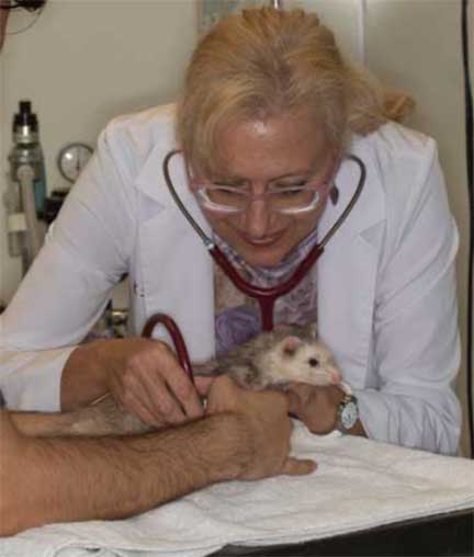
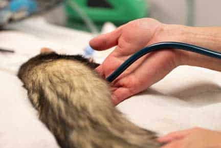
We use a special stethoscope (called an esophageal stethoscope) that is passed down the esophagus and can give us a clear sound of the heart
Intestinal Foreign Body Surgical Removal
The two different foreign bodies were removed surgically. Both ferrets recovered completely within a few days after surgery.

Our patient is carefully clipped and scrubbed
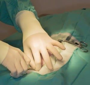
Our surgeon does the all important draping after the final cleanse of the abdomen

Under these drapes is a hot water blanket and also hot water bottles

When our surgeon is comfortable everything is in order he makes his skin incision

There is a special location on the abdominal muscles called the linea alba. You can see it as this white horizontal line between the muscles.
It is here that our surgeon cuts through the muscle and enters the abdominal cavity without cutting any abdominal muscles. This is a tendon that holds the abdominal muscles together, and is the area that has the best holding strength when we suture the area back together. If it did not have this strength, we would have an abdominal hernia.

The scissors is used to make the cut through the linea alba into the abdomen
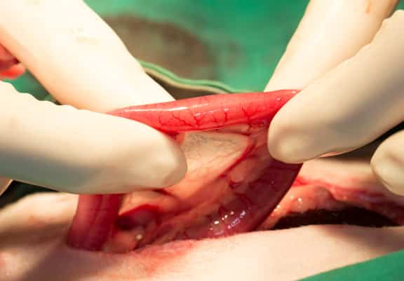
Even though the surgeon knows the location of the foreign body (duodenum) from the ultrasound, he palpates every inch of the intestines to make sure nothing is abnormal or missed.
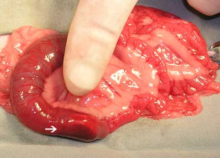
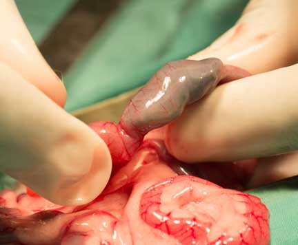
The white arrow on the bottom points to the circular foreign body. Note how distended the intestine is to the left of the arrow. This is that ileus seen on the radiograph. Also note how the distended intestine is hemorrhagic compared to the normal and not inflamed intestines on the right.
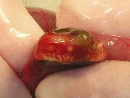
Our surgeon localizes the foreign body and packs the area with gauze before incising into it and removing the FB
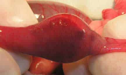
Another view of the FB and how stuck it is
Intestine incised and FB removed
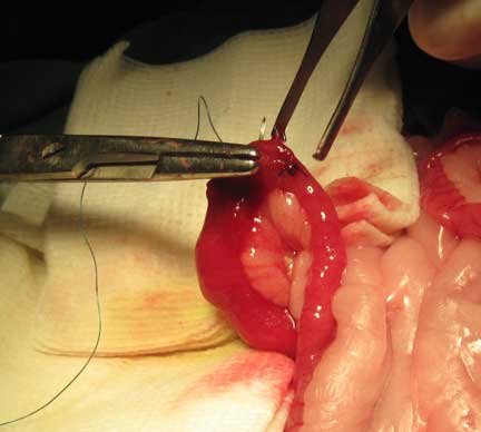
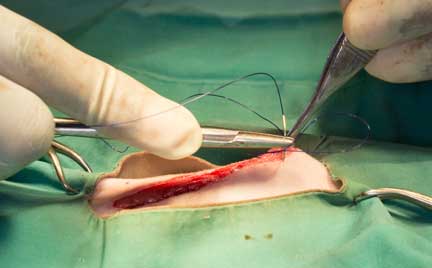
Suturing the opening in the intestine is crucial, and our surgeon gently and carefully closes the open with suture material that will dissolve over several weeks
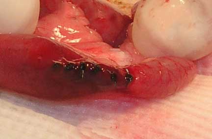
The final closure of the intestines with what is called a “simple interrupted” suture pattern
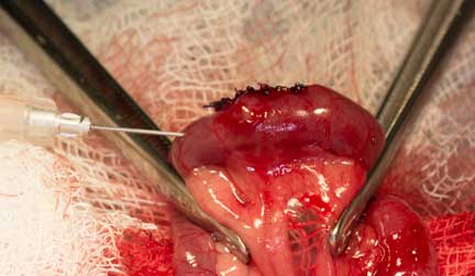
A tiny needle with sterile saline is gently injected into the area to make sure there are no leaks before finishing the procedure.
When our surgeon is satisfied the intestines will not leak the linea alba, the all important tendon between the abdominal muscles, is carefully sutured.
This is what the plastic foreign body looksed like in the small intestines
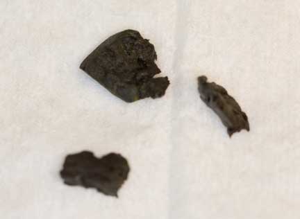
There were several pieces
On every exploratory surgery, once the initial problem is addressed, we check all the internal organs to make sure there are no other problems.
This is the spleen
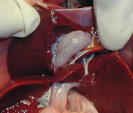
The liver with the gallbladder in the center
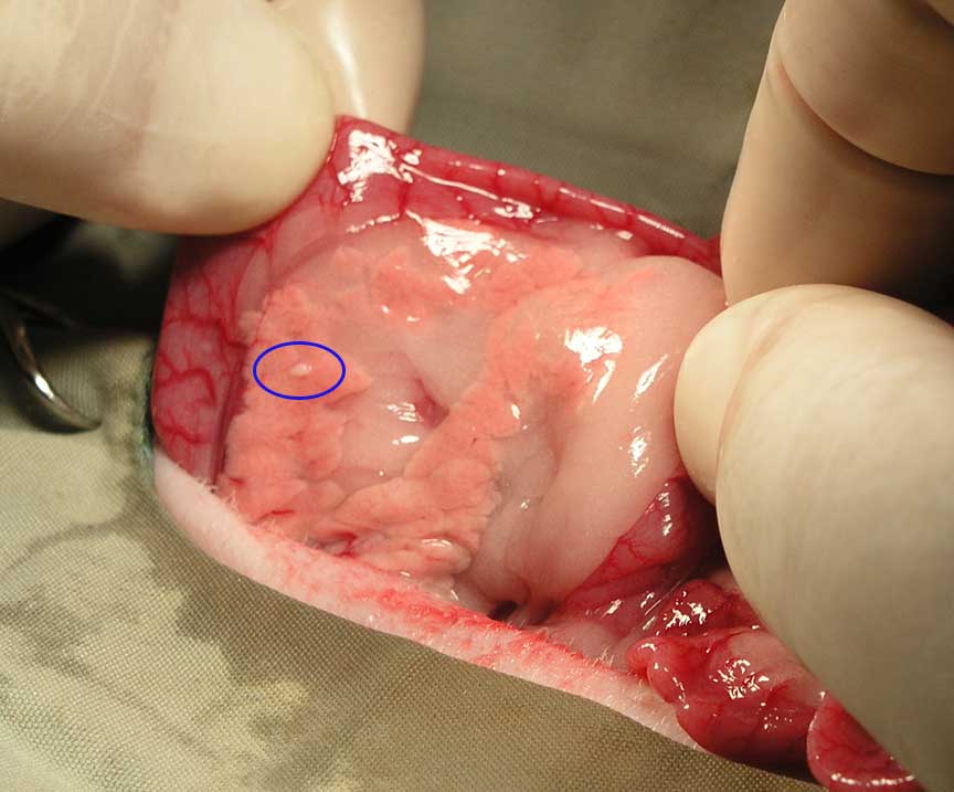
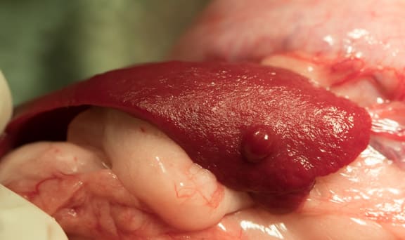
Checking the pancreas. The blue circled area is an insulinoma nodule.
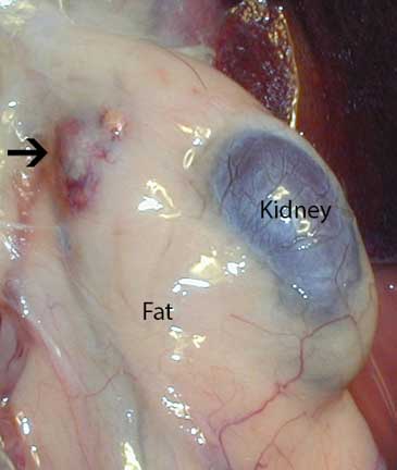
The left kidney and an inflamed left adrenal gland buried in fat. This is typical of Adrenal Gland disease.
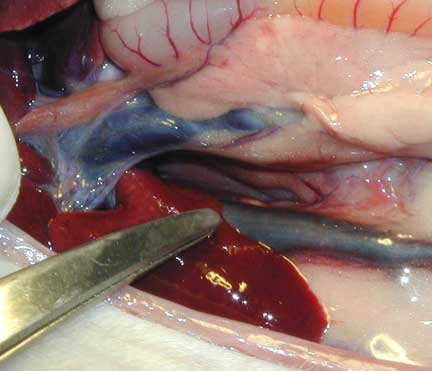
Below the scissors is a lobe of the liver. Horizontally at the very top is the small intestines. The horizontal dark blue object is the vena cava, the vessel that returns venous blood from the abdominal organs back to the right side of the heart.
Post-Operative Care

Our surgery patients get therapy laser treatment to minimize pain and aid in the healing process
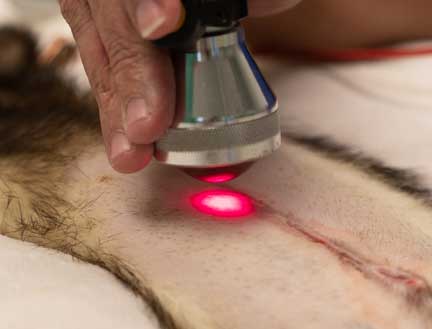
The laser penetrates the skin at the incision and works on the tissue below
At this point in the surgery a pain injection will be given and the patient allowed to wake up slowly.
After surgery it is off to recovery
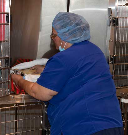
While in recovery it is monitored closely by the nursing staff
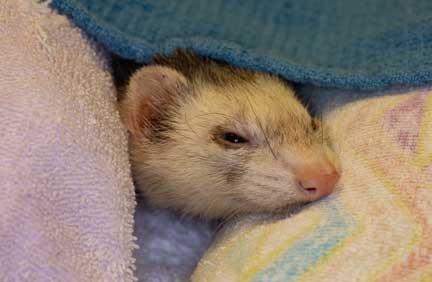
Our patient cozy and warm in her blanket, comfortably resting after surgery
Time to go home after an eventful day
Student Externs
We are a registered teaching hospital that has trained many veterinary students over the last 35 years in our Externship Program. Getting to be a part of a ferret spay is invaluable to them since they do not get this exposure in veterinary school. The work they do with us is hands-on, and starts them on their way to getting the skills necessary to work with ferrets.
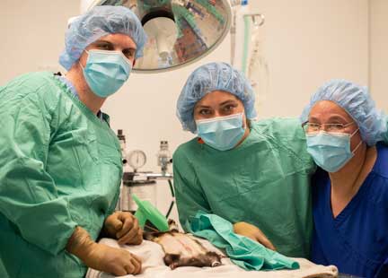
Our extern David Harris from the U. C. Davis veterinary school with our surgical team
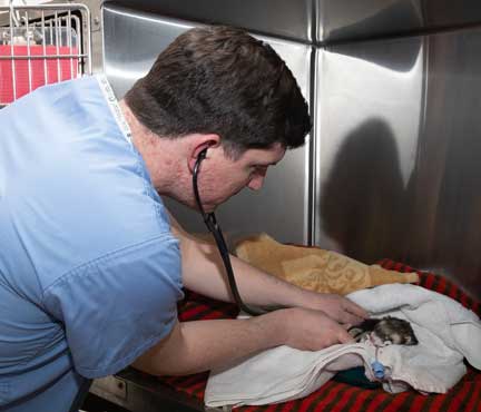
David learning how to monitor our ferret spay patient in recovery
Our externs are required to post their daily experience at our hospital on our Facebook page. It is called the Extern Daily Diary. If you go to our Facebook page and scroll down for many years you will see the many posts they have written.
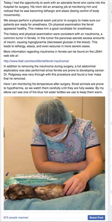
This student made her Facebook post on a ferret that had an insulinoma surgery
Ferrets make wonderful pets. They are prone to several diseases; the two most important you need to be aware of if you want to have a ferret for a pet are: